Leg Bone Diagram - This illustration shows the normal skeletal structure of ... - Master leg and knee anatomy using our topic page.. The humerus and the femur are corresponding bones of the arms and legs, respectively. Pngtree offers bone diagram png and vector images, as well as transparant background bone diagram clipart images and psd files. Femur bone indicated in purple. Each leg is made up of four bones. He'll boost his body knowledge as he matches up the names of the bones with their proper places on the leg diagram.
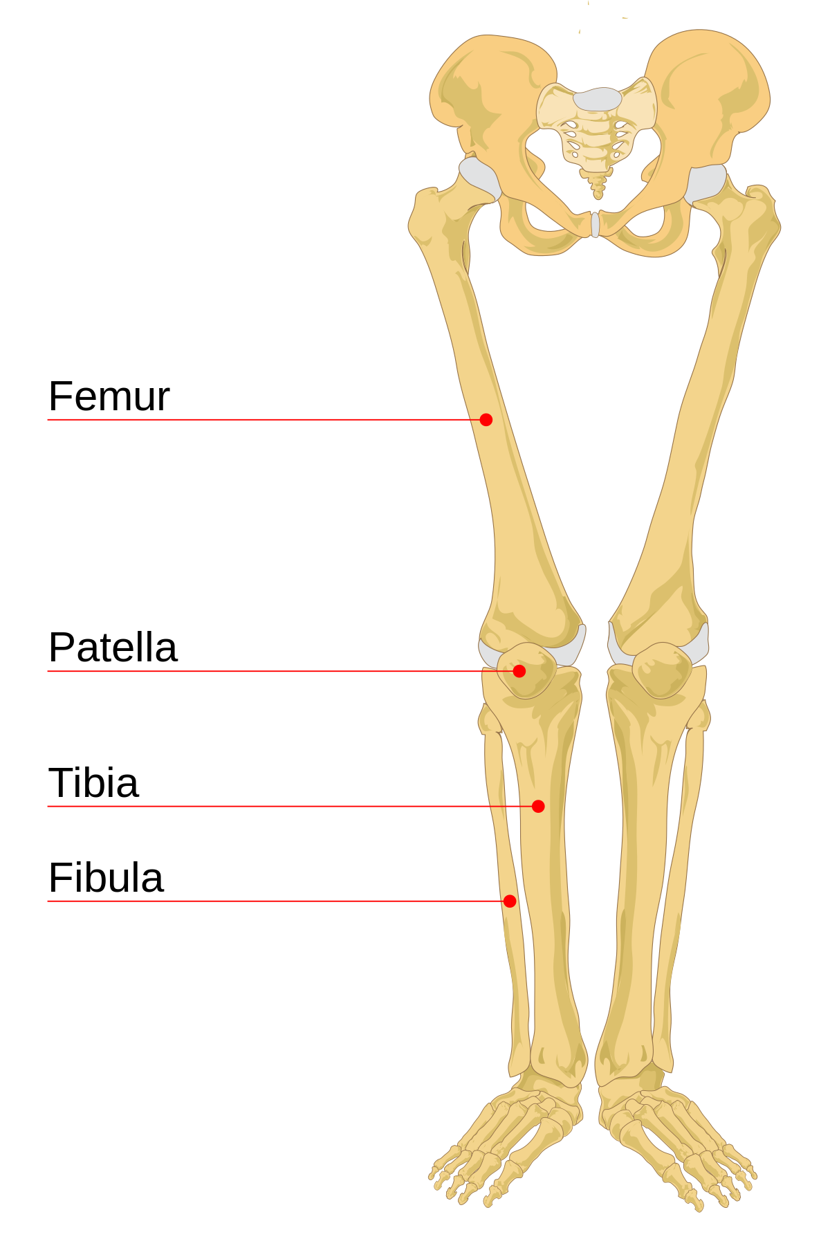
25.09.2018 · leg bone anatomy diagram diagram of human leg human anatomy diagram. In this image, you will find femur, medial epicondyle of the femur, patella, tibial tuberosity, anterior rest of. The tibia is the main bone of the leg, forming what is more commonly known as the shin. This bright worksheet helps your child bring these technical terms down to size. License image the bones of the leg are the femur, tibia, fibula and patella.
Bones of the Human Leg and Foot - ScienceAid from scienceaid.net High quality realistic skeleton legs. Click now to learn more about the bones, muscles, and soft tissues tibia: The tibia is the main bone of the leg, forming what is more commonly known as the shin. In this image, you will find femur, medial epicondyle of the femur, patella, tibial tuberosity, anterior rest of. Learn vocabulary, terms and more with flashcards, games and other study tools. Your legs are two of your most important body parts. Each leg is made up of four bones. Femur bone indicated in purple.
The foot bones shown in this diagram are the talus, navicular, cuneiform, cuboid, metatarsals.
Time to jump right into the biggest and strongest bones in the human body. Pngtree offers bone diagram png and vector images, as well as transparant background bone diagram clipart images and psd files. Includes leg (femur, tibia, patella, and fibula) and foot (tarsals and digits) bones. The radius and ulna (bones of the forearm), shown in supination (the arm rotated outward so that the palm. New users enjoy 60% off. This long bone connects with the knee at one end and the next to the tibia is the fibula, the thinner, weaker bone of the lower leg. Learn how to draw the femur, patella, tibia, and fibula in this lesson! In this image, you will find femur, medial epicondyle of the femur, patella, tibial tuberosity, anterior rest of. The foot bones shown in this diagram are the talus, navicular, cuneiform, cuboid, metatarsals and calcaneus. They allow you to move and provide support for your upper body. The bones of the leg are the femur, tibia, fibula and patella. At the same time, the bones and joints of the leg and foot must be strong enough to support the body's weight while remaining flexible enough for movement and balance. Start studying leg bone diagram.
Pngtree offers bone diagram png and vector images, as well as transparant background bone diagram clipart images and psd files. He'll boost his body knowledge as he matches up the names of the bones with their proper places on the leg diagram. Master leg and knee anatomy using our topic page. Start studying leg bone diagram. Explore the fascination world of human bones.
Exam 2 Bones of the Lower Limb - Anatomy 329 with ... from s3.amazonaws.com Learn how to draw the femur, patella, tibia, and fibula in this lesson! In the leg, the interosseous membrane extends between the tibia and the fibula, running along the crests of the bones. Download 2,751 bone diagram stock illustrations, vectors & clipart for free or amazingly low rates! Your leg bones are the longest and strongest bones in your body. This long bone connects with the knee at one end and the next to the tibia is the fibula, the thinner, weaker bone of the lower leg. Download the free graphic resources in the form of png, eps. Master leg and knee anatomy using our topic page. The largest and most medial leg bone, forming both the knee and ankle joints.
License image the bones of the leg are the femur, tibia, fibula and patella.
Your legs are two of your most important body parts. Learn how to draw the femur, patella, tibia, and fibula in this lesson! The humerus and the femur are corresponding bones of the arms and legs, respectively. New users enjoy 60% off. The largest and most medial leg bone, forming both the knee and ankle joints. Lower jaw (mandible) collar bone. Cheek bone (zygoma) upper jaw (maxilla). Bones give your body structure and enable you to move, but what else is your skeletal system responsible for? The red bone marrow inside of bones produces most of the blood cells, including erythrocytes (red blood cells), leukocytes (white blood cells), and thrombocytes (platelets). When you stand or walk, all the weight of your upper body rests on them. Your leg bones are the longest and strongest bones in your body. The foot bones shown in this diagram are the talus, navicular, cuneiform, cuboid, metatarsals and calcaneus. Quizzes on human skeletal system anatomy, bone anatomy, and bone markings.
This long bone connects with the knee at one end and the next to the tibia is the fibula, the thinner, weaker bone of the lower leg. Each leg is made up of four bones. Includes leg (femur, tibia, patella, and fibula) and foot (tarsals and digits) bones. They allow you to move and provide support for your upper body. The largest and most medial leg bone, forming both the knee and ankle joints.
Leg bone - Wikipedia from upload.wikimedia.org New users enjoy 60% off. The tibia is the main bone of the leg, forming what is more commonly known as the shin. Quizzes on human skeletal system anatomy, bone anatomy, and bone markings. Start studying leg bone diagram. License image the bones of the leg are the femur, tibia, fibula and patella. Lower jaw (mandible) collar bone. Explore the fascination world of human bones. Time to jump right into the biggest and strongest bones in the human body.
Your legs are two of your most important body parts.
You'll learn about the muscles, bones, and other structures of each area of the leg. New users enjoy 60% off. Download the free graphic resources in the form of png, eps. At the same time, the bones and joints of the leg and foot must be strong enough to support the body's weight while remaining flexible enough for movement and balance. Femur bone indicated in purple. Cheek bone (zygoma) upper jaw (maxilla). Includes leg (femur, tibia, patella, and fibula) and foot (tarsals and digits) bones. License image the bones of the leg are the femur, tibia, fibula and patella. The bones of the leg are the femur, tibia, fibula and patella. The tibia is the main bone of the leg, forming what is more commonly known as the shin. Master leg and knee anatomy using our topic page. High resolution textures and displacement included. The foot bones shown in this diagram are the talus, navicular, cuneiform, cuboid, metatarsals.
Berbagi :
Posting Komentar
untuk "Leg Bone Diagram - This illustration shows the normal skeletal structure of ... - Master leg and knee anatomy using our topic page."


Posting Komentar untuk "Leg Bone Diagram - This illustration shows the normal skeletal structure of ... - Master leg and knee anatomy using our topic page."